


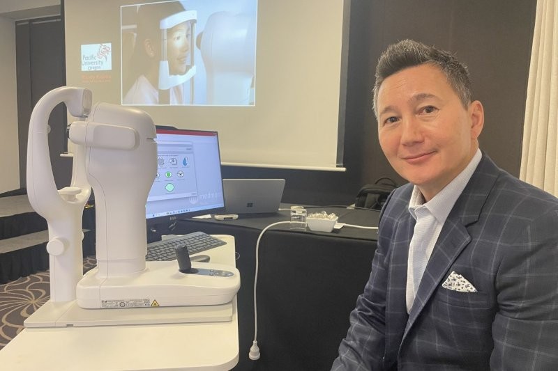
Distributed in New Zealand by Ophthalmic Instrument Company, the Medmont Meridia Vantage is being positioned as a comprehensive anterior eye imaging platform, with scleral topography its key advance.
Speaking at the recent CCLS conference, Randy Kojima, specialty contact lens practitioner and R&D collaborator at Precision Technology Services, Vancouver, Canada, introduced the device as a step towards more data-driven scleral lens fitting.
“There’s a new Medmont that does scleral topography and that’s what the team asked me to share with you,” he said. “We are actually measuring the sclera, so it should improve the efficiency and accuracy of scleral-lens fitting.”
The instrument captures scleral shape using fluorescein reflection, either in a single shot or by merging up to five images into a composite map. In more challenging cases, composite capture can extend coverage to approximately 20mm. Kojima noted that, worldwide, approximately 90% of patients are fitted with a 16–17mm or smaller scleral lens, but the additional diameter measurements may assist with deep-set eyes or when selecting scleral lenses larger than 17mm.
Accuracy is still being evaluated. “I’d love to tell you that this thing is accurate to 10µ, but we really don’t know yet,” Kojima said. Early indications suggest results are “usually within about 50µ of where we think it is” for diagnostic lens selection, with some internal estimates closer to 15µ, under ideal conditions.
A non-fluorescein scleral mapping mode is expected within six months. “This will be an option to take corneal topography with the Placido and do a non-fluorescein scan of the sclera so you can merge the two together,” Kojima said. The approach is intended to assist patients who drain dye rapidly and to provide combined corneal, limbal and scleral analysis without fluorescein.
He described the contact lens software as enabling practitioners to place a theoretical lens on a measured ocular surface, rather than estimating scleral shape. “It’s kind of exciting we have that tool,” he said. “It’s not only an orthokeratology tool, but one that can help fit virtually any lens, from soft contact lenses to scleral lenses, all with the same device.”
Beyond scleral mapping, Meridia Vantage incorporates corneal topography, white-light imaging, infrared meibography and dry-eye analysis. Kojima said the aim is to support initial lens selection, clinical documentation and patient education using a single high-resolution system with dual light sources to minimise shadowing.
“So we can now fit scleral lenses with a level of accuracy in both diagnostic fitting and custom lenses not previously possible,” he said.