


Researchers in Japan found microneedles as small as 34G may be suited to biopsies of intraocular fluids and suspicious lesions, potentially leaving self-healing wounds.
Writing in Clinical & Experimental Ophthalmology, researchers from Yokohama City University Medical Centre said they assessed microneedles of different sizes in lab models, including puncturing PVC.
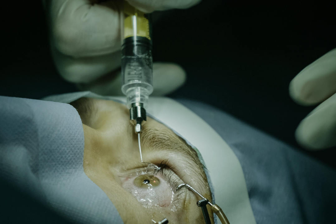
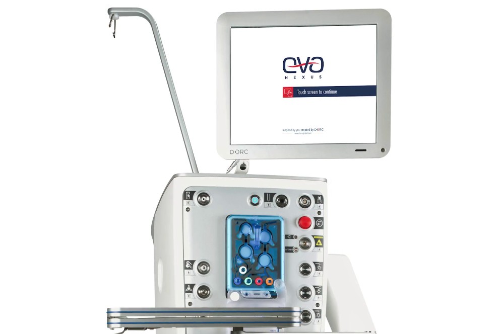
Retinal microneedles, defined as having at least one dimension between 1 and 999μm, have been developed to cannulate retinal vasculature with the goal of treating vaso-occlusive diseases, said the research team. “Such microneedles can be coupled with vitrectomy machines, such as the Alcon Constellation Vision System, and have an outer diameter of 50μm and an estimated lumen of 35μm. The microneedle therefore has a 8.26, 6.24 and 2.22 times smaller outer diameter than the 27G, 30G and 38/41G polytip, respectively, which may result in smaller retinotomy and less invasive means to retrieve biopsy material.”
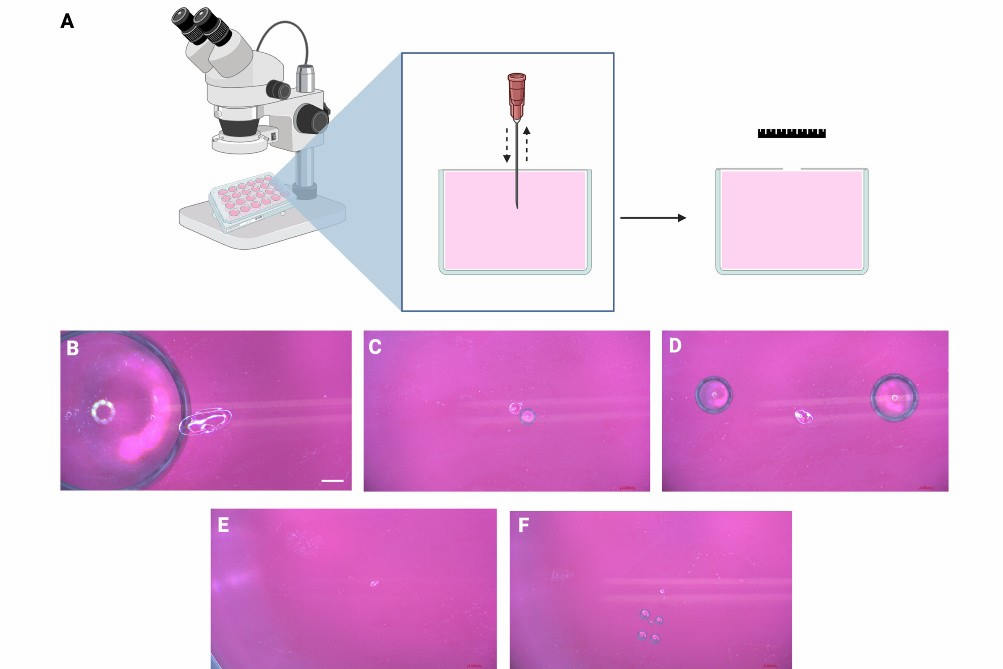
The study’s assessments comprised: human uveal melanoma retinoblastoma cancer cell lines being aspirated using microneedles with a vitrectomy setup and cell viability analysis; suspensions of cancer cells with fluorescent microbeads being injected into the subretinal space of fresh ex-vivo porcine eyes before simulating biopsy with microneedle retinal puncture, followed by imaging with OCT and histology; and anterior chamber puncture performed with microneedles and imaged with anterior segment OCT and examined for aqueous leakage.
Authors said instrument choice should consider precise dimensional characteristics to balance minimal trauma with sufficient tissue sampling and warrants further in-vivo studies in animal models.